Pulmonary Manifestation among Patients with Systemic Lupus Erythematosus (SLE) in Khartoum State 2022
Ziryab Mahmoud¹², Alaa Osman³, Tarig Ahmed¹²
Keywords:
Systemic lupus erythematosus, Pulmonary manifestations, Interstitial lung disease, Pulmonary fibrosis, Pleural effusionAbstract
Background: Systemic lupus erythematosus (SLE) is a chronic, multisystem autoimmune inflammatory disease that predominantly affects young women. Pulmonary involvement is variably reported across studies and ranges from mild pleural disease to interstitial lung disease (ILD), with pleural effusion and pulmonary fibrosis among the commonly described abnormalities.
Objective: To determine the prevalence and pattern of lung manifestations among patients with SLE.
Methods: A multicenter, observational, descriptive cross-sectional study was conducted in rheumatology clinics in Khartoum State, Sudan, during 2022. All patients diagnosed with SLE according to the 2019 classification criteria were included. Data were collected, cleaned, and analyzed using SPSS version 25.0.
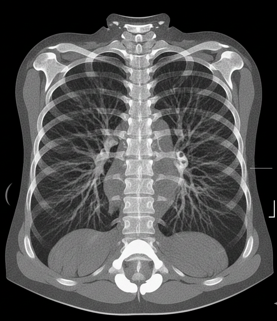
Results: The study included 53 patients with SLE, with a mean age of 39.3 ± 12.6 years and a male-to-female ratio of 1:52. Approximately half of participants (27, 50.9%) had been diagnosed with SLE for 1–5 years. The most frequent presenting symptoms were fever (14, 26.4%), cough (14, 26.4%), shortness of breath (10, 18.9%), and chest pain (7, 13.2%). Symptom duration of less than one year was reported by 26 patients (49.1%). Common comorbidities included hypertension (9, 17%), heart disease (3, 5.7%), and diabetes mellitus (2, 3.8%). Laboratory findings showed ANA positivity in 52 patients (98.1%); anti–dsDNA positivity in 23 (43.4%); elevated CRP in 12 (22.6%); elevated ESR in 33 (62.3%); low complement C3 in 8 (15.1%); and low complement C4 in 11 (20.8%). On chest X-ray, pleural effusion was noted in 2 (3.8%), lung fibrosis in 1 (1.9%), and reticulonodular shadowing in 1 (1.9%). On chest CT, ILD/pulmonary fibrosis was the most frequent finding (10, 18.9%), followed by pleural effusion (4, 7.5%); pulmonary nodule (1, 1.9%); nonspecific findings (1, 1.9%); ascites (1, 1.9%); and patchy consolidation (1, 1.9%). Echocardiography was normal in 9 patients (17%); ischemic heart disease was identified in 2 (3.8%); diastolic dysfunction in 1 (1.9%); mild aortic and tricuspid regurgitation in 1 (1.9%); and pulmonary hypertension in 1 (1.9%). Regarding treatment, most participants received hydroxychloroquine (47, 88.7%), followed by azathioprine (15, 28.3%), mycophenolate mofetil/Cellcept (11, 20.8%), prednisone (10, 18.9%), and leflunomide (1, 1.9%).
Conclusion: Pulmonary manifestations among Sudanese patients with SLE were clinically relevant and should not be overlooked, particularly ILD/pulmonary fibrosis and pleural effusion. These findings support strengthening screening, early detection, and targeted prevention and management policies for high-risk SLE patients in Sudan.
Downloads